- 1.1.1 The objectives and development of cell biology
- 1.2.1 Microscope and Cell theory
- 1.3.1 Major events in cell biology
- 2.1.1 cell culture
- 2.2.1 Monoclonal antibodies
- 2.3.1 Fluorescence Microscopy
- 2.4.1 Flow cytometry
- 2.5.1 Spinning Disk Confocal Microscopy
- 2.6.1 FRAP and FRET
- 2.7.1 RNA interference
- 3.1.1 The structrures of the plasmic membrane
- 3.2.1 The lipid bilayer Composition and structural organization
- 3.3.1 Forms of lipid molecules exist in water
- 3.4.1 Lipids Movement in biomembrane
- 3.5.1 Membrane Proteins
- 3.6.1 Synthesis of Phospholipids and Intracellular Movement
- 5.1.1 Overview of transmembrane transport
- 5.2.1 Facilitated Transport of Glucose and Water
- 5.3.1 ATP –powered pumps
- 5.4.1 Na+k+ pump
- 5.5.1 Ca2+ pump
- 5.6.1 K+ channel and the Resting membrane potencial
- 5.7.1 Cotransport by Symporters and Antiporters
- 5.8.1 Transcellular transport
- 6.1.1 Overview of aerobic oxidation
- 6.2.1 Structure of Mitochondria
- 6.3.1 Mitochondrial Oxidation of Fatty Acids Generates ATP
- 6.4.1 The Electron Carriers and Electron Transport Chain
- 6.5.1 The Electron Transport and Proton Motive Force
- 6.6.1 Chemiosmotic theory and Endosymbiont hypothesis
- 6.7.1 Harnessing the Proton-Motive Force to synthesize ATP
- 6.8.1 Electron Transport and Reactive Oxygen Species (ROS)
- 7.1.1 Targeting Proteins to and Across the ER Membrane
- 7.2.1 Insertion of single-pass Proteins into the ER
- 7.3.1 Insertion of Multipass Proteins into the ER
- 7.4.1 Protein Glycosylation and Folding in the ER
- 7.5.1 Formation of disulfide bonds in the ER
- 7.6.1 Protein Quality Control in the ER
- 7.7.1 Targeting of Proteins to Mitochondria
- 7.8.1 Targeting of Peroxisomal Proteins
- 8.1.1 Molecular Mechanisms of Vesicle Budding and Fusion
- 8.2.1 Early Stages of the Secretory Pathway
- 8.3.1 Later Stages of the Secretory Pathway
- 8.4.1 Receptor-Mediated Endocytosis
- 8.5.1 Directing Membrane Proteins to the Lysosome
- 8.6.1 The structure and functions of Lysosomes
- 9.1.1 Brief Introduction of Cell Communication
- 9.2.1 Signaling Molecules and Receptors
- 9.3.1 G Proteins and G Protein-coupled Receptors (GPCR)
- 9.4.1 GPCR Mediated Signaling pathways
- 10.1.1 Outlines of Signaling pathways that control gene activity
- 10.2.1 Phosphoinositide Signaling Pathways
- 10.3.1 TGF-b Receptors and TGFb Signaling Pathways
- 10.4.1 Signaling Pathways controlled by Ubiquitination
- 10.5.1 The RasMAPK and JAKSTAT signaling pathway
- 11.1.1 Actin Filaments
- 11.1.2 Actin Filaments
- 11.2.1 Dynamic Assembly of Actin Filaments
- 11.3.1 Actin-Based Cellular Structures
- 11.4.1 Myosins and Myosin-Powered Movements
- 12.1.1 Microtubule Structure and Organization
- 12.2.1 Microtubule Dynamics
- 12.3.1 Regulation of Microtubule Structure and Dynamics
- 12.4.1 Kinesins & Dyneins Microtubule-Based Motor Proteins
- 12.5.1 Cilia & Flagella Microtubule-Based Surface Structures
- 12.6.1 Mitosis & Mitotic Spindle
- 12.7.1 Intermediate Filaments
- 12.8.1 Coordination & Cooperation Between Cytoskeletal Elements
- 13.1.1 Nuclear Envelope Nuclear Pore Complex & Nuclear Transport
- 13.2.1 Structural Organization of Eukaryotic Chromosomes
- 13.3.1 Morphology and Functional Elements of Eukaryotic Chromosomes
- 13.4.1 Nucleolus
- 14.1.1 Unit 1 Overview of the cell cycle and its control
- 14.2.1 Unit 2 Model Organisms and Methods to Study the Cell Cycle
- 14.3.1 Unit 3 CDK and Its Discovery
- 14.4.1 Unit 4 Regulation of CDK Activity
- 14.5.1 Unit 5 Commitment to the Cell Cycle and DNA Replication
- 14.6.1 Unit 6 Entry into Mitosis and Completion of Mitosis
- 14.7.1 Unit 7 Surveillance Mechanisms in Cell Cycle Regulation
- 14.8.1 Unit 8 Meiosis A Special Type of Cell Division
- 15.1.1 An Overview on Cell-Cell and Cell-Matrix Adhesion
- 15.2.1 Type of Cell Junctions and Anchoring Junctions
- 15.3.1 Tight junctions and Gap junctions
- 15.4.1 The Extracellular Matrix I_the Basal Lamina
- 15.5.1 ECMs of Connective tissue
- 16.1.1 Early metazoan developmnt and embryonic stem cells
- 16.2.1 Stem Cells and Niches in Multicellular Organisms
- 16.3.1 Mechanisms of cell polarity & asymmetric cell division
- 16.4.1 Cell death and its regulation
探索生命的最小单位:厦门大学细胞生物学深度解读课
如果你对生命如何运转充满好奇,这门细胞生物学课程绝对是打开微观世界大门的钥匙。
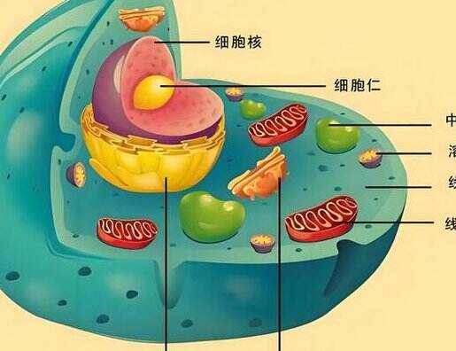
从显微镜到分子机制的科学之旅
细胞生物学可不是简单的背背名词。厦门大学这门课最吸引人的地方在于,它带我们穿越三个观察维度——用显微镜看细胞全貌,用电子显微镜探索亚细胞结构,最后深入到分子水平理解运作原理。
教授们特别注重把枯燥的理论变成看得见的实验。比如讲到细胞膜运输时,会带大家做荧光标记实验,亲眼观察物质是如何穿过细胞膜的。这样的实操环节让抽象概念变得触手可及。
课程亮点抢先看
生物膜系统的神奇之处
你知道吗?细胞膜不只是简单的"包装纸"。我们将详细拆解:为什么有些物质能自由进出细胞,有些却需要特殊通道?能量转换细胞器如何像微型发电厂一样工作?
细胞骨架:细胞的"钢筋水泥"
微丝、微管这些听起来高大上的结构,其实就像细胞内部的脚手架。课程会用三维动画演示它们如何支撑细胞形态、参与细胞运动。
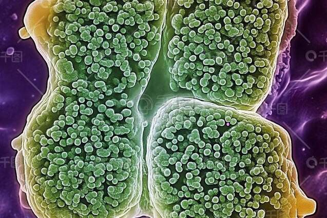
遗传信息的精密管理
从DNA到蛋白质的完整生产线是怎样的?细胞核这个"指挥部"如何确保遗传信息精准传递?这部分内容会和分子生物学知识紧密衔接。
适合哪些人学习?
- 生物相关专业本科生打基础
- 准备考研的同学系统复习
- 对生命科学感兴趣的跨专业学习者
- 需要更新知识的在职科研人员
特别值得一提的是,课程精心设计了从基础到前沿的渐进式学习路径。即使没有太多生物学背景,也能通过生动的案例教学跟上进度。
完整课程模块一览
开篇导论:从细胞学说的建立到现代技术突破
实验方法专题:单克隆抗体技术、荧光显微镜实操、流式细胞术解析
膜系统深度剖析:磷脂双分子层的动态特性、膜蛋白的合成与组装
物质运输专场:钠钾泵的工作机制、协同运输的分子原理
能量工厂探秘:线粒体的超微结构、ATP合成中的质子动力
学完能达到什么水平?
- 能够解释细胞结构与功能的关系
- 掌握主流细胞生物学研究技术
- 理解癌症、干细胞等热点问题的细胞学基础
- 具备阅读细胞生物学前沿论文的基本能力
厦门大学的这门细胞生物学课,不仅教授知识,更培养科研思维。每次课后的专题讨论都值得期待,可以和同学一起探讨最新发表的细胞生物学发现。